| |
Anterior Knee Pain (Kneecap)
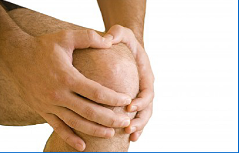
Anterior knee pain is a broad term used to describe pain felt at the front of the knee, around or behind the kneecap (patella). It is a symptom rather than a single diagnosis, and it may arise from several different problems affecting the kneecap and the knee extensor mechanism.
Common causes include patellofemoral pain due to overuse or muscle imbalance, irritation or degeneration of the patellar tendon below the kneecap, quadriceps tendinopathy above the kneecap, patellar instability where the kneecap does not track properly or partially slips out of place, and arthritis affecting the joint between the kneecap and the thigh bone. Less commonly, anterior knee pain may also follow an injury to the extensor mechanism, which includes the quadriceps muscle, quadriceps tendon, patella, patellar tendon, and their attachment to the shin bone.
Symptoms
The symptoms vary depending on the underlying cause, but patients commonly complain of pain at the front of the knee during walking, running, squatting, kneeling, climbing or descending stairs, and getting up from a chair. The pain may also be felt after sitting for a prolonged period with the knee bent.
In tendon-related problems such as patellar or quadriceps tendinopathy, the pain is usually localised either just below or just above the kneecap and is often worse during jumping, sprinting, deep bending, or repeated loading activities. In patellar instability, patients may describe a sense of the kneecap slipping, shifting, giving way, or even dislocating. Arthritis may cause pain together with stiffness, grinding, swelling, and difficulty with daily activities. If the extensor mechanism is significantly injured, there may be marked weakness or an inability to actively straighten the knee.
Diagnosis
The diagnosis starts with a detailed history and specialist clinical examination. This helps determine the site of the pain, whether the symptoms are related to overuse, instability, wear and tear, or an acute injury, and whether there are any mechanical symptoms such as catching, locking, or recurrent giving way.
On examination, the knee will be assessed for tenderness, swelling, range of movement, patellar tracking, ligament stability, muscle strength, and the integrity of the extensor mechanism. The ability to perform a straight leg raise is particularly important when assessing more significant tendon or patellar injuries.
Investigations may include X-rays to assess the kneecap position, alignment, and any arthritic change. MRI scans can be helpful to evaluate the cartilage, tendons, soft tissues, and associated injuries. In selected cases, ultrasound may also be useful, particularly for tendon-related conditions.
Treatment
Treatment depends entirely on the underlying cause, the severity of the symptoms, and the patient’s activity level.
Many cases can be managed without surgery. Non-operative treatment may include activity modification, pain relief medication, ice, physiotherapy, muscle strengthening, stretching, and correction of poor lower limb mechanics. Physiotherapy is particularly important to improve quadriceps strength, hip control, flexibility, and patellar tracking. Bracing, taping, or a patellar support may also help in selected cases.
For patellar or quadriceps tendinopathy, treatment is usually based on a structured rehabilitation programme with gradual tendon loading and temporary reduction of aggravating activities such as jumping and running. For patellar instability, a first episode may often be treated with rest, bracing, and rehabilitation, while recurrent instability may require surgical stabilisation. In patellofemoral arthritis, treatment may include physiotherapy, pain relief, injections, and activity modification.
Further detailed information and the appropriate treatment options will be provided by Mr. Ayoub during the consultation. Back...
|
|